This is an abnormal ECG with T wave inversion in V1-3. A troponin done next, was significantly elevated. The diagnosis is MYOCARDITIS.
- Arrhythmias: Beware the febrile child with sinus tachycardia, where the rate is out of proportion to the fever. There may be premature atrial or ventricular beats, supraventricular tachycardias and even ventricular tachycardias. Bradycardias including complete heart block can also occur(2,3)
////////////////////////////
MYOCARDITIS
Myocarditis is simply an inflammation of the heart muscle, that can lead to myocardial cell damage, myocardial dysfunction and heart failure.
A Recent Case
A 15 yo male presents to the emergency department with sharp central chest pain that is worst on inspiration. The pain has woken him from sleep. There is no radiation although he states “My left arm feels strange”.
He has no significant past medical history, but has had a recent episode of fevers and rigors, with a frontal headache and a sore throat. The local doctor has commenced Amoxicillin for a diagnosis of sinusitis.
He is treated by the overnight registrar with Paracetamol and Ibuprofen and his chest pain improves.
Clinically he looks well, is afebrile and has dual hear sounds, with no added sounds, a clear chest on auscultation and a soft abdomen.
He has a normal chest xray and his bloods show a WCC 12.9, Neutrophils of 8.88 and a CRP of 68
His ECG is shown below. What do you think?
 Using the ECG in 20 Seconds Method we can quickly say the following:
Using the ECG in 20 Seconds Method we can quickly say the following:- HR = 90bpm
- P axis is normal= sinus rhythm
- P’s correspond to QRS’s
- QRS is not too tall or small, not too wide, and doesn’t have abnormal morphology
- ST-T segments show abnormal T wave inversion in V1-V3( perhaps a little biphasic looking)
- PR and QT intervals look normal
- There are no pacing spikes- not that you would expect there to be.
Is this T wave inversion normal? We know that in the first week of life the T waves are upright in the anterior leads. They then flip and stay that way until adolescence(usually age 10-12 years) Therefore these are abnormal. (Read more on paediatric ECG’s).
A troponin(not high sensitivity) was done that was 8.5 (<0.04). This 15 yo had a clinical working diagnosis of myocarditis.
How do patients with myocarditis present?
The presentation is variable in both children and adults and ranges from subclinical disease to arrhythmias, cariogenic shock and death. We need to have a high clinical suspicion for the disease.
It can present as simple fatigue, impaired exercise tolerance and respiratory distress. When in fulminant myocarditis, children will present with haemodynamic compromise, which may lead to cardiovascular collapse. Children are more likely to present with acute or fulminant disease, but may present with respiratory or gastrointestinal symptoms, that can lead to an incorrect diagnosis. Missing the diagnosis can be potentially disastrous, with a percentage of children progressing to dilated cardiomyopathy and requiring cardiac transplant.
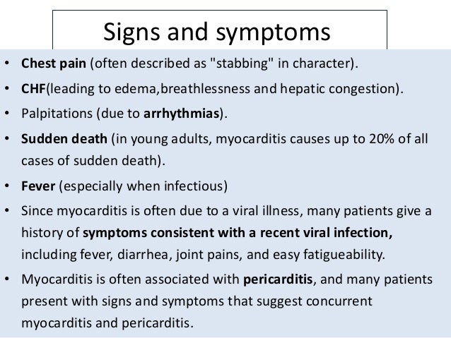
The main presenting symptoms are:
- Chest Pain: This may mimic myocardial ischaemia, especially in younger children(1). It may simply be the pain of pericarditis. In those with pericarditis and a troponin leak, the diagnosis is myopericardits and is secondary to epicardial inflammation. Patients can also present with symptoms of pericarditis, ie sharp, sudden onset of chest pain that is aggravated by inspiration and coughing. These patients may have Perimyocarditis.
- Respiratory Distress: Patients may present with dyspnoea at rest and tachypnoea, which in children, can lead to a mistaken diagnosis of respiratory disease, such as chest infection or bronchiolitis or asthma.
- Abdominal Symptoms: Children can present with abdominal pain and vomiting, leading to a misdiagnosis of gastrointestinal-related disease.
- Arrhythmias: Beware the febrile child with sinus tachycardia, where the rate is out of proportion to the fever. There may be premature atrial or ventricular beats, supraventricular tachycardias and even ventricular tachycardias. Bradycardias including complete heart block can also occur(2,3)
How Common Is It?
The annual incidence in children is very small, less than 1 in 100,00 per annum. We know that this is an underestimate as we do not pick up subclinical disease, which is an issue, as it recurs in a percentage of these children. The concerning findings in respect to its incidence, is that in autopsies of children, that have died unexpectedly, myocarditis is found in about 20%(4,5).
What Causes It?
The most common cause in children is viral. This includes enterovirus, adenovirus, Epstein Barr virus and others(6).It can also be caused much less commonly by auto-immune disorders and drug hypersensitivity. The primarily viral aetiology make it very important to note a recent history of respiratory, or gastrointestinal illness.
How Do We Make The Diagnosis?
History and Examination
A high clinical suspicion is paramount, especially in patients who present with viral-like illnesses and symptoms such as myalgia, fatigue, but also with chest pain, respiratory distress, unexplained sinus tachycardia or new onset of heart failure. In children we must be suspicious of respiratory illness, on the background of a viral prodrome.
We must suspect it in patients with the following findings(6):
- Cardiac Dysfunction
- Elevated Cardiac Markers
- An abnormal ECG
- An Echocardiogram showing ventricular dysfunction with no structural cause found.
The clinical examination may reveal signs of cardiac failure, especially left ventricular failure, extra heart sounds(an S3 or S4) and hepatomegaly.
Investigations
ECG
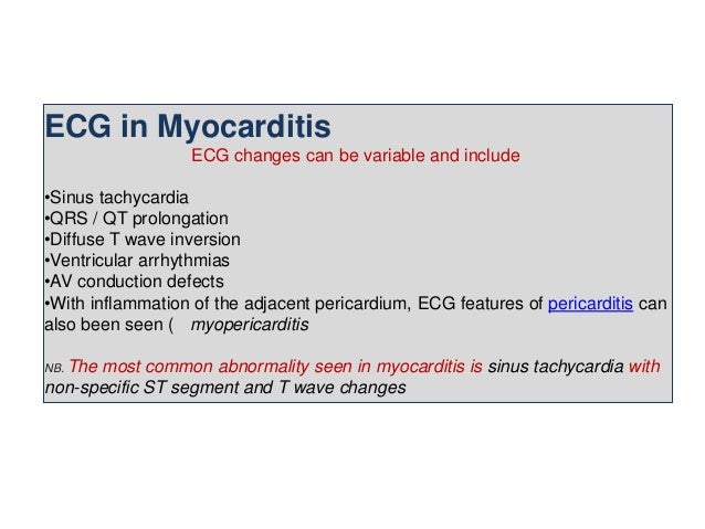
The ECG is abnormal, however there are no particularly sensitive ECG findings(7,8). Changes that can occur include:
- ST-T segment abnormalities including T wave inversion
- Atrial or ventricular enlargement
- Arrhythmias including:
- sinus tachycardia
- premature atrial and ventricular beats
- supraventricular tachycardia
- ventricular tachycardia
- complete heart block
Cardiac Markers
These include troponin, but may not always be elevated. If there is a suspicion of heart failure a BNP should also be done.
Chest Xray
It has limited sensitivity, but may show cardiomegaly, pulmonary venous congestion or pleural effusions. In children it may be important to rule out a pneumonia especially if the presentation is that of fever and respiratory distress.
Echocardiography
The echocardiogram can detect changes in left ventricular function, even in situations when the disease is subclinical(9). It can also demonstrate changes in ventricular shape and size, wall motion abnormalities, pericardial involvement and valvular abnormalities.
Endomyocardial Biopsy
This is considered the gold standard for making the diagnosis, however it has a poor sensitivity, at best 50%(10). There procedure also has a risk of cardiac wall perforation, especially in children.
Cardiac MRI
Although biopsy is considered to be the gold standard, we see cardiac MRI, being used with increasing frequency. It can identify the location and extent of inflammation and its diagnostic accuracy approaches 80% in adults, although, we are less certain of its sensitivity in children.
Do adult patients need cardiac catheterisation?
This is not usually required, except for situations where myocarditis cannot be distinguished from a potential ischaemic cause.
Summary
A high degree of clinical suspicion should exercised especially if patients have an abnormal ECG and abnormal cardiac markers. In children the presentation of a recent viral prodrome and respiratory distress or even gastrointestinal symptoms, with nothing to find, we should be suspicious of an underlying myocarditis. The incidence is low, however that is the very reason we should think of it.
References
- Mahrholdt H, Wagner A, Deluigi CC, et al. Presentation, patterns of myocardial damage, and clinical course of viral myocarditis. Circulation 2006; 114:1581.
Myocarditis is a condition resulting from inflammation of the heart muscle that presents with a broad clinical spectrum of signs and symptoms in affected children ranging from subclinical disease to cardiogenic shock (ie, fulminant myocarditis), arrhythmias, and sudden death (table 2). (See 'Clinical manifestations' above.)
●Myocarditis should be suspected in a child with signs and symptoms of cardiac dysfunction including respiratory distress (table 2), a rise in cardiac biomarkers (eg, troponin), electrocardiographic changes suggestive of acute myocardial injury or arrhythmia, and/or echocardiographic evidence of cardiac dysfunction without an underlying structural cardiac defect. Chest radiography findings are nonspecific and include cardiomegaly, pulmonary vascular congestion, and, less commonly, pleural effusions (image 1 and image 2); however, about half of affected patients have normal chest radiographs. In patients with suggestive findings, it is desirable to perform further testing (eg, endomyocardial biopsy [EMB] and cardiac magnetic resonance imaging [MRI]) that may confirm the diagnosis of myocarditis. However, these tests may not be readily available or may be inconclusive, in which case management decisions are made based on a clinical diagnosis that encompasses these findings. (See 'Clinical manifestations' above and 'Clinical diagnosis' above.)
●EMB is considered the gold standard to confirm a clinical diagnosis of myocarditis; however, the sensitivity of this test is low and the test requires invasive cardiac catheterization. Nevertheless, in our clinical practice, we typically perform endomyocardial biopsy in patients with clinically suspected myocarditis to confirm the diagnosis. We focus on performing EMB early in the course of the disease and obtaining multiple samples to optimize diagnostic accuracy and reduce sampling errors. The diagnosis of myocarditis from EMB is made based on standard light microscopy (Dallas), immunohistochemistry, and viral genomic analysis criteria. (See 'Endomyocardial biopsy' above.)
●Cardiac MRI is increasingly used to confirm the diagnosis in children with myocarditis using consensus criteria based on studies in adults. Cardiac MRI can document areas of cardiac inflammation. In addition to EMB, we also perform cardiac MRI in the majority of our patients. (See 'Magnetic resonance imaging' above.)
●Further diagnostic evaluation is performed to determine the underlying etiology of myocarditis. This includes viral cultures from rectal and nasal mucosal swabs and antibody titers, and evaluation based on signs/symptoms indicative of systemic diseases including Lyme disease, acute rheumatic fever, or systemic lupus erythematosus. (See 'Further diagnostic evaluation' above.)
●The differential diagnosis of myocarditis includes other diseases that present with respiratory distress (eg, pneumonia) orsigns/symptoms of heart failure (eg, hypertrophic cardiomyopathy). These conditions usually can be distinguished from myocarditis by history, examination, and laboratory tests. (See 'Differential diagnosis' above.)
No comments:
Post a Comment