

CERVICAL LYMPH NODE DISSECTION.
14TH FEBRUARY 2015
Lymph Nodes draining tissues of the head and neck are located in
the fibrofatty tissue that lies between
the superficial (investing)layer of the deep fascia and
the deeper visceral and prevertebral layers .
the fibrofatty tissue that lies between
the superficial (investing)layer of the deep fascia and
the deeper visceral and prevertebral layers .
In this space the nodes lie aggregated around certain neural and vascular structures such as
1- Internal jugular vein,
2-Spinal accessory nerve, and
3- Transverse cervical artery.
4- Trachea and Throid -(not included in Radical or Modified Radical Neck dissections)
1- Internal jugular vein,
2-Spinal accessory nerve, and
3- Transverse cervical artery.
4- Trachea and Throid -(not included in Radical or Modified Radical Neck dissections)
Much about the details of the anatomy (location) and the drainage zones of these nodes have been elucidated
First through Lymhangiography studies done in the past three decades.–and later on through image based studies using CTScans and MRI.
Based on these studies the earlier anatomic classification of these nodes initially got redefined into 5 categories: namely -
Junctional, Jugular,(superficial and Deep) Spinal, Supraclavicular, and Retroauricular.
However, the nomenclature in popular use today comes from the study of the patterns of metastatic dissemination observed in patients who were treated at the center with radical neck dissection. (The Memorial Sloan Kettering Cancer Center studies).
In this nomenclature, Lymph nodes in the neck are grouped into levels I-V,
Level 1- Submental and Submandibular nodes.
Levels II, III, IV - Upper, Middle, and Lower jugular nodes ;
Level V -Posterior triangle nodes
Level VI – Midline of the Neck
In this nomenclature, Lymph nodes in the neck are grouped into levels I-V,
Level 1- Submental and Submandibular nodes.
Levels II, III, IV - Upper, Middle, and Lower jugular nodes ;
Level V -Posterior triangle nodes
Level VI – Midline of the Neck
The 6 levels of the neck nodes with sublevels.
 |
| Ref Medscape |
Level I
Boundaries-
Superiorly -The lower border of theBody of the mandible ,
Inferiorly -A line running horizontally from the inferior border of the hyoid bone.
Anteriorly -The anterior belly of the digastric muscle on the contralateral side .
Posteriorly -the vertical plane marked by the posterior edge of the submandibular gland is used as a boundary between levels I and II. -(2008) ----
The earlier posterior boundary was The stylohyoid muscle,
Subdivisions
Level I may be divided into
Level I may be divided into
Level Ia, which refers to the nodes in the submental triangle (bound by the anterior bellies of the digastric muscles and the hyoid bone), and
Level Ib, which refers to the submandibular triangle nodes (see image ).
Level Ib, which refers to the submandibular triangle nodes (see image ).
 |
| www.pixshark.com |
Closely related, to the level Ib group of nodes, are The perifacial nodes, related to the facial vessels above the mandibular margin, and The buccinator nodes, (see image below)
Drainage areas -
The nodes of level Ia drain -
The floor of mouth, / Anterior tongue,/ Anterior mandibular alveolar ridge, and Lower lip
The nodes of Level Ib - The oral cavity, / Anterior nasal cavity/, Skin and soft tissue structures of the midface, and Submandibular gland.
The prefacial and Buccinator nodes may become involved with metastasis from tumors in the
Buccal mucosa,/ Nose, and Skin and Soft tissues of the cheek and lips -
 |
| www.studyblue.com |
The nodes of level Ia drain -
The floor of mouth, / Anterior tongue,/ Anterior mandibular alveolar ridge, and Lower lip
The nodes of Level Ib - The oral cavity, / Anterior nasal cavity/, Skin and soft tissue structures of the midface, and Submandibular gland.
The prefacial and Buccinator nodes may become involved with metastasis from tumors in the
Buccal mucosa,/ Nose, and Skin and Soft tissues of the cheek and lips -
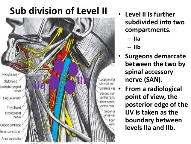
Level II
Boundaries Extend from
The skull base Superiorly to Inferiorly the inferior border of the hyoid bone
They are related to the upper third of the jugular vein,.
The anterior border of level II is the vertical plane marked by the posterior edge of the submandibular gland
The 2008 classification revision proposed that the vertical plane marked by the posterior edge of the submandibular gland be used as an alternative to the stylohyoid muscle boundary
The posterior border of level II is the posterior border of the sternocleidomastoid muscle,
 |
| www.pocketdentistry.com |
Subdivisions
The spinal accessory nerve, which travels obliquely across this area, is used as a landmark to subdivide this group
IIa, the part that lies anteroinferiorly and closer to the internal jugular vein
IIb, the portion above and behind the nerve,

The spinal accessory nerve, which travels obliquely across this area, is used as a landmark to subdivide this group
IIa, the part that lies anteroinferiorly and closer to the internal jugular vein
IIb, the portion above and behind the nerve,
Drainage areas - . The oral cavity,/ Nasal cavity,/ Nasopharynx, Oropharynx, Hypopharynx, Larynx, and Parotid gland. (see image below)
 |
| www.vocalclinic.org |
Level III
Boundaries
Superiorly The Hyoid Bone
Inferiorly -a horizontal plane defined by the inferior border of the cricoid cartilage. This line also traverses the OmoHyoid muscle at this level.
Anterior border -The sternohyoid muscle
Inferiorly -a horizontal plane defined by the inferior border of the cricoid cartilage. This line also traverses the OmoHyoid muscle at this level.
Anterior border -The sternohyoid muscle
The posterior border is posterior border of the sternocleidomastoid muscle .
Subdivisions -Nil
The nodes at this level lie around the middle third of the Internal Jugular vein.
The nodes at this level lie around the middle third of the Internal Jugular vein.
Drainage areas - Oral cavity, Nasopharynx, Oropharynx, Hypopharynx, and Larynx.-
(see image below)
(see image below)
 |
| www.medicalgeek.com |
Level IV
Boundaries -
Superior boundary – Inferior border of the Cricoid cartilage-
Inferior boundary -The Clavicle.
Anterior boundary -SternoHyoid muscle.
Posterior boundary -Posterior border of the Sternomastoid muscle.
Inferior boundary -The Clavicle.
Anterior boundary -SternoHyoid muscle.
Posterior boundary -Posterior border of the Sternomastoid muscle.
These nodes at this level lie in relation to the lower third of the jugular vein.
Subdivisions -Nil.
Drainage areas - The larynx, hypopharynx, thyroid, and cervical esophagus as shown below.
 |
| www.kuleuven.be |
Level V
This refers to the lymph nodes located in the posterior triangle of the neck.(see image below)
 |
| www.rahulgladwin.com |
Boundaries
Superior boundary - The apex of the convergence of the sternocleidomastoid and trapezius muscle
Inferior boundary – Clavicle.
Anterior Boundary - Posterior border of the Sternomastoid muscle
Posterior boundary - The anterior border of the Trapezius muscle
Inferior boundary – Clavicle.
Anterior Boundary - Posterior border of the Sternomastoid muscle
Posterior boundary - The anterior border of the Trapezius muscle
Subdivisions -subdivided by a plane defined by the inferior border of the cricoid cartilage into
level Va superiorly and
level Vb inferiorly.
level Va superiorly and
level Vb inferiorly.
Level Va contains the nodes associated with the Spinal accessory nerve, and
Level Vb contains the Transverse cervical and Supraclavicular nodes.
Level Vb contains the Transverse cervical and Supraclavicular nodes.
Drainage areas - The nasopharynx, oropharynx, and skin of the posterior scalp and neck.
The Supraclavicular nodes connect with the Mediastinal group of Nodes
The Supraclavicular nodes connect with the Mediastinal group of Nodes
Level VI
This refers to lymph nodes of the anterior, or central, compartment of the neck.
Boundaries
Superiorly – The hyoid bone.
Inferiorly – The Suprasternal notch.
Laterally – Carotid arteries.
 |
| www.youtube.com |
Inferiorly – The Suprasternal notch.
Laterally – Carotid arteries.
Subdivisions -Nil
Drainage areas
Thyroid gland,
Subglottic larynx,
Cervical trachea,
Hypopharynx, and
Cervical esophagus .
Subglottic larynx,
Cervical trachea,
Hypopharynx, and
Cervical esophagus .
Lymph nodes in this compartment are located in
1-The tracheoesophageal groove (paratracheal nodes),
2-In front of the trachea (pretracheal nodes),
3-Around the thyroid gland (Para-Thyroidal nodes), and
4-On the cricothyroid membrane (Precricoid or Delphian node).
(Level VII- Mediastinum)
1-The tracheoesophageal groove (paratracheal nodes),
2-In front of the trachea (pretracheal nodes),
3-Around the thyroid gland (Para-Thyroidal nodes), and
4-On the cricothyroid membrane (Precricoid or Delphian node).
 |
| www.surgicalcore.org |
Summary of drainage areas of Cervical Lymph Nodes.
 |
No comments:
Post a Comment